Multiple Sclerosis Normal Cervical Spine Mri
Imaging Of The Spine In Multiple Sclerosis Practice Essentials
Imaging Of The Spine In Multiple Sclerosis Practice Essentials

Imaging In Multiple Sclerosis Journal Of Neurology Neurosurgery
The Radiology Assistant Diagnosis And Differential Diagnosis

Evaluation Of Focal Cervical Spinal Cord Lesions In Multiple

Multiple Sclerosis Cervical Spinal Cord Clinical Mri

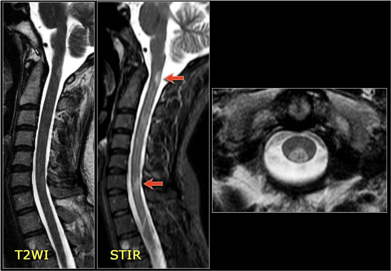
Lesions are usually the most telling.
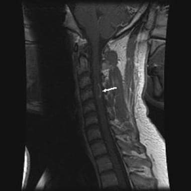
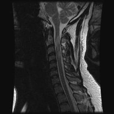
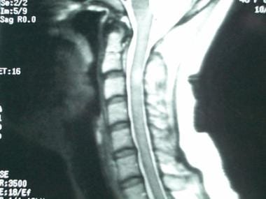
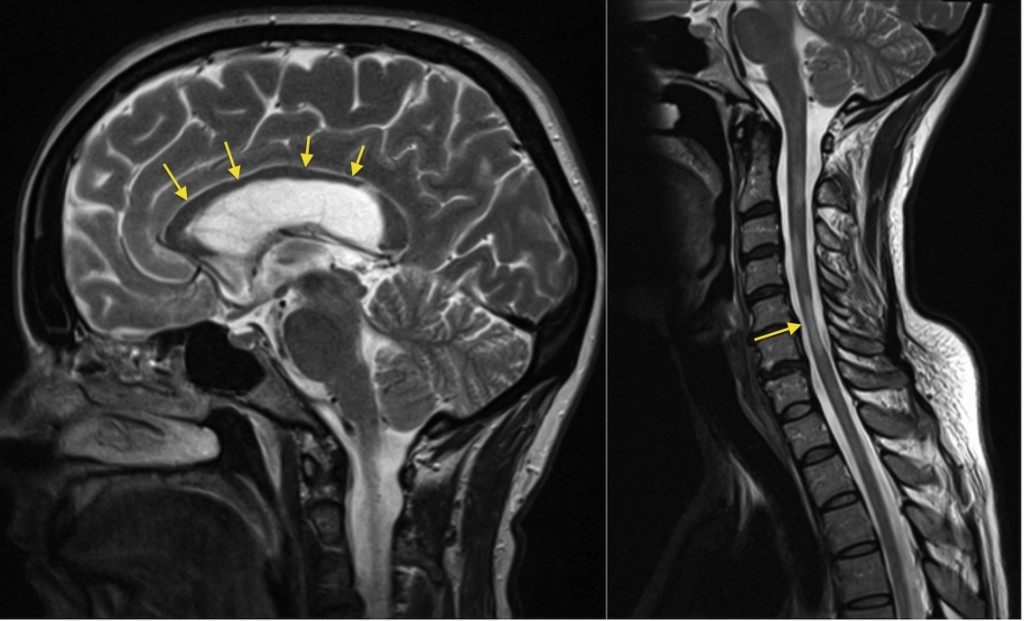
Multiple sclerosis normal cervical spine mri. Disease related changes in the brain or spinal cord are detected by. Spinal ms lesions often occur in the cervical region and less frequently in the lower thoracic spinal cord t7 12 depending on their age ms plaques appear normal or slightly hypointense on t1 weighted images and hyperintense on t2. An mri result that says things are normal does not absolutely rule out multiple sclerosis. Practice essentials magnetic resonance imaging mri was first used to visualize multiple sclerosis ms in the upper cervical spine in the late 1980s.
About 5 of people with multiple sclerosis don t have lesions in the brain that show up on the test. The most effective and non invasive way to determine if a person has ms is to scan for brain and spinal cord lesions using magnetic resonance imaging mri. The spinal cord may be enlarged when the disease is active and is atrophied when chronic. Ms care team widespread use of mri magnetic resonance imaging has revolutionized the ability to diagnose multiple sclerosis.
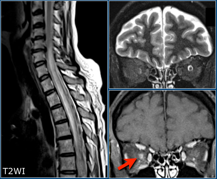
Mri stands for magnetic resonance imaging mri can reveal telltale areas of damage called lesions or plaques on. 1 s spinal ms is often associated with. Multiple sclerosis ms is a relatively common acquired chronic relapsing demyelinating disease involving the central nervous system and is the second most common cause of neurological impairment in young adults after trauma 19 characteristically and by definition multiple sclerosis is disseminated not only in space i e. In multiple sclerosis ms by definition lesions are separated in space and time.
A type of imaging test called an mri scan is an important tool in diagnosing ms. Multiple lesions in different regions of the brain but also in time.
Imaging Of The Spine In Multiple Sclerosis Practice Essentials

Spinal Cord Abnormalities In Recently Diagnosed Ms Patients

Science Source Multiple Sclerosis Of Cervical Spinal Cord

Multiple Sclerosis Cervical And Thoracic Cord Lesions Clinical Mri

Evaluation Of Focal Cervical Spinal Cord Lesions In Multiple
Multiple Sclerosis Spinal Cord Atrophy Radiology At St

Double Inversion Recovery Sequence Of The Cervical Spinal Cord In

Healthy Cervical Spine Mri

Sample Images Mri Defined Spinal Cord Lesion Versus Normal
The Radiology Assistant Myelopathy
Thoracic Spinal Cord Lesions Are Influenced By The Degree Of

Multiple Sclerosis Cervical Spinal Cord Clinical Mri

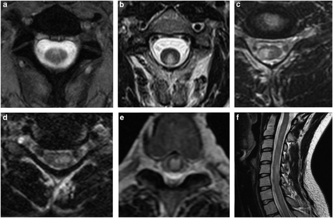
Fig 1 Comparison Of Three Mr Sequences For The Detection Of